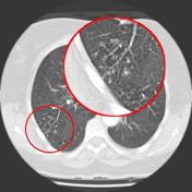
tree in bud opacities pneumonia
Usually somewhat nodular in appearance the tree-in-bud pattern is generally most pronounced in the lung periphery and associated with abnormalities of the larger airways. These findings are intriguing.
Tib opacities are also associated with bronchiectasis and small airways obliteration resulting in mosaic air trapping.
. Cryptogenic organizing pneumonia 119. While the tree-in-bud appearance usually represents an endobronchial spread of infection given the proximity of small pulmonary arteries and small airways sharing branching morphology in the bronchovascular bundle a rarer cause of the tree-in-bud sign is infiltration of the small pulmonary arteriesarterioles or. Cytomegalovirus pneumonia in a 51-year-old man with chronic myelogenous leukemia who underwent bone marrow transplantation.
Note the scattered lung nodules surrounded by. More extensive lympho - cytic infiltrations may be associated with lymphoid interstitial pneumonia LIP with ground-. Here Patel and colleagues showed an association between the presence of the vascular tree-in-bud pattern yes vs.
No and duration of ventilation and hospitalization at the time of CT imaging with a majority of patients who demonstrated vascular tree-in-bud on CT imaging experiencing 10 or more days of ventilation and hospitalization both P 001. The purpose of this study was to determine the relative frequency of. TIB opacities represent a normally invisible branches of the bronchiole tree 1 mm in diameter that are severely impacted with mucous pus or fluid with resultant dilatation and budding of the terminal bronchioles 2 mm in diameter 1 photo.
Foreign material entering into the tracheobronchial tree secondary to gastroesophageal reflux altered mental status drug overdose anesthesia or neurologic disorder stroke traumatic brain injury Often develops in intubated. Jennifer hong ba francisca zuazo md hanyuan shi md 1 1 tulane university la new orleans. Franquet T et al.
The tree-in-bud pattern can be an early sign of disease Fig 10 15. Thrombotic microangiopathy of pulmonary tumors. Tree-in-bud pattern at thin-section CT of the lungs.
Pneumonia due to respiratory syncytial virus in a 23-year-old man with leukemia. Aspiration Pneumonia and Tree in Bud Sign 87 year old male with history of cough and suspicion of aspiration shows barium aspiration into the proximal trachea upper right The scout view upper right shows an infiltrate at the right base Thickened airways in the right lower lobe 2nd row left is associated with a pneumonic infiltrate in the right lower lobe lower right. Although initially described in 1993 as a thin-section chest CT finding in active tuberculosis TIB opacities are by.
What does tree-in-bud opacities mean. Patients with aspiration pneumonia are some-times complicated with Mycobacterium infections especially elderly patients. We investigated the pathological basis of the tree-in-bud lesion by reviewing the pathological specimens of bronchograms of normal lungs and contract radiographs of the post-mortem lungs manifesting active pulmonary.
This is the classic appearance of the tree in bud pattern seen on chest ct. Nodules 110 mm in diameter may be seen in a number of lung infections. These small clustered branching and nodular opacities represent terminal airway mucous impaction with adjacent peribronchiolar inflammation.
We here describe an unusual cause of TIB during the COVID-19 pandemic. TIB opacities represent a normally invisible branches of the bronchiole tree 1 mm in diameter that are severely impacted with mucous pus or fluid with resultant dilatation and budding of the terminal bronchioles 2 mm in diameter1 photo. Tree-In-Bud Pattern A lymphoid interstitial infiltrate in the walls of the small airways follicular bronchiolitis may cause small centrilobular nodules and the tree-in-bud pattern Fig.
Are tree-in-bud nodules cancerous. It is most commonly associated with infectious diseases affecting the bronchioles1 OP resulting in a tree in bud pattern has been previously suggested2 However a clear radiological-pathological correlation of OP filling the bronchioles resulting in a tree in bud pattern has to the best of our knowledge not yet been clearly demonstrated. Multiple causes for tree-in-bud TIB opacities have been reported.
When physicians discover a tree-in-bud pattern the patient should be placed in a negative pressure room immediately. However to our knowledge the relative frequencies of the causes have not been evaluated. Rossi SE et al.
1012 Poorly defined centrilobular nodules associated with branching linear and nodular opacities ie tree-in-bud sign are the typical HRCT findings of infective bronchiolitis frequently. And tree-in-bud branching opacities detected throughout both lung fields after aspiration. Thin-section CT scan shows peripheral poorly defined centrilobular nodules and tree-in-bud opacities bilaterally.
AJR Am J Roentgenol. Nodules micronodules and tree-in-bud opacities. Tree-in-bud TIB opacities are a common imaging finding on thoracic CT scan.
A Thin-section CT scan of the right lung shows centrilobular ground-glass opacities in addition to nodules and tree-in-bud opacities arrow. An 82yearold man was transferred to our emergency department with symptoms of chills and 405C fever. Treeinbud opacities detected after aspiration should be considered DAB rather than mycobacterial infection.
Although initially described in 1993 as a thin-section chest CT finding in active tuberculosis TIB opacities are by. HR-CT patterns seen in OP are. Can pneumonia cause lung opacity.
A young male patient who had a history of fever cough and respiratory distress presented in the emergency department. Tree-in-bud TIB appearance in computed tomography CT chest is most commonly a manifestation of infection. The purpose of this study was to determine the relative frequency of causes of TIB opacities and identify patterns of disease associated with TIB opacities.
Radiology scientific expert review panel. In the acute phase bacterial pneumonia manifests in the form of segmental or lobar consolidation Fig 2 possibly with cavitation and related hilar and mediastinal adenopathies. Tree-in-bud refers to a pattern seen on thin-section chest CT in which centrilobular bronchial dilatation and filling by mucus pus or fluid resembles a budding tree.
Lobular ground-glass opacity may be seen in patients with infection eg bronchopneumonia viral infections Pneumocystis jirovecii pneumonia or M pneumoniae. Since the initial report of endobronchial spread of pulmonary tuberculosis the tree-in-bud sign has been reported in a wide variety of health conditions including infectious diseases aspiration pneumonia congenital disorders idiopathic disorders inhalation immunologic disorders connective disorders 23456 and central lung cancer involving the. A vascular cause of tree-in-bud pattern on CT.

References In Causes And Imaging Patterns Of Tree In Bud Opacities Chest

Pdf Tree In Bud Semantic Scholar

Tree In Bud Sign And Bronchiectasis Radiology Case Radiopaedia Org

Hrct Scan Of The Chest Showing Diffuse Micronodules And Tree In Bud Download Scientific Diagram

Tree In Bud Pattern Pulmonary Tb Eurorad

References In Causes And Imaging Patterns Of Tree In Bud Opacities Chest

Tree In Bud Sign Lung Radiology Reference Article Radiopaedia Org

Tree In Bud Pattern Radiology Case Radiopaedia Org
View Of Tree In Bud The Southwest Respiratory And Critical Care Chronicles
View Of Tree In Bud The Southwest Respiratory And Critical Care Chronicles

Tree In Bud Pattern Radiology Case Radiopaedia Org

Chest Ct With Multifocal Tree In Bud Opacities Diffuse Bronchiectasis Download Scientific Diagram

References In Causes And Imaging Patterns Of Tree In Bud Opacities Chest
